Pathological Fluid Segmentation in OCT with U-Net
Ablation study on retinal fluid segmentation in OCT images comparing six U-Net configurations. Best configuration (ResNet34 + BCE+Dice): Dice 0.824, IoU 0.716, AUC-ROC 0.993 on 5-fold cross-validation.
Visit websiteResearch Context
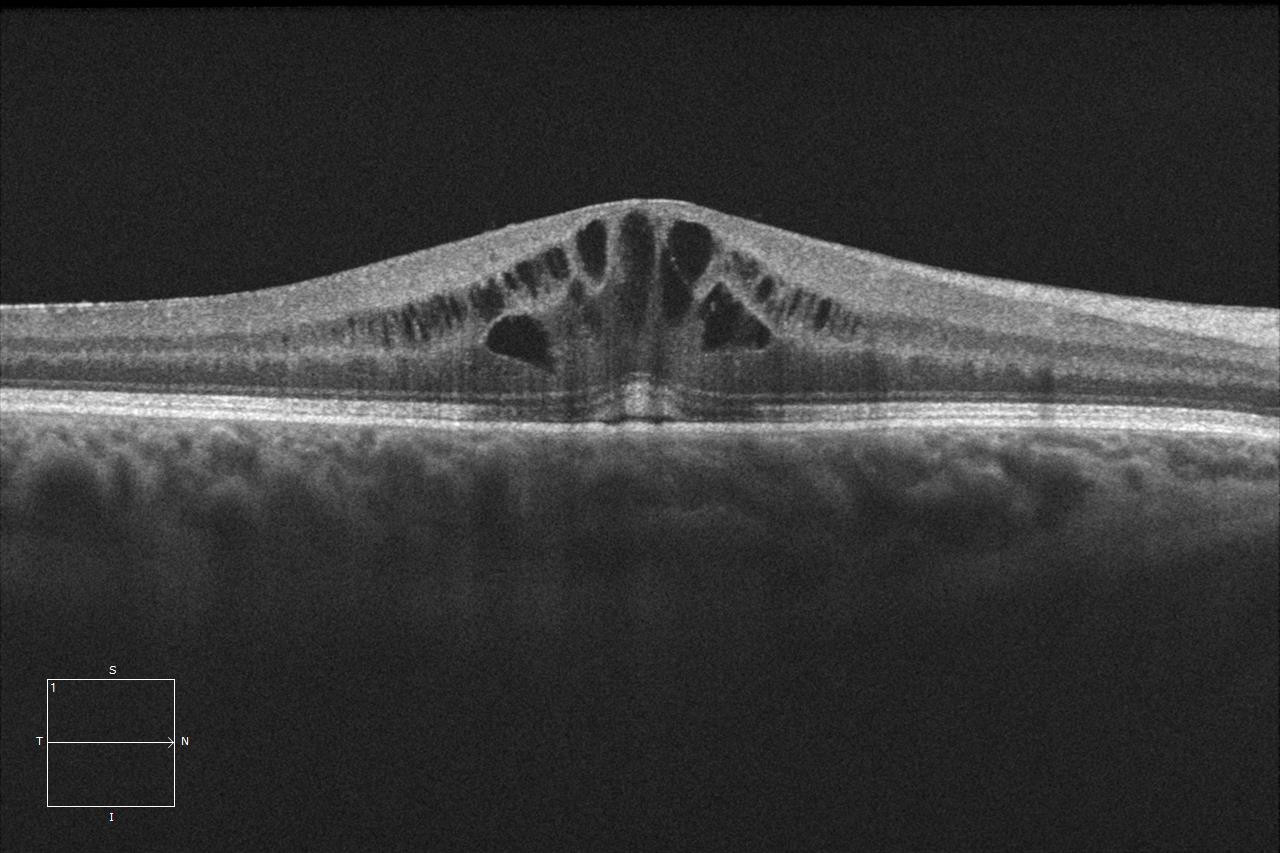
Optical Coherence Tomography (OCT) is the gold standard for retinal diagnosis, but manual segmentation of pathological fluid (DME, SRF, PED) is time-consuming and observer-dependent. Automated pixel-level segmentation enables faster, reproducible clinical decisions.
Ablation Study — Results
| Configuration | Dice | IoU | AUC-ROC |
|---|---|---|---|
| Scratch + BCE | 0.741 | 0.609 | 0.981 |
| Scratch + Dice | 0.763 | 0.635 | 0.984 |
| ResNet34 + BCE | 0.798 | 0.672 | 0.989 |
| ResNet34 + Dice | 0.811 | 0.695 | 0.991 |
| ResNet34 + BCE+Dice ★ | 0.824 | 0.716 | 0.993 |
| ResNet34 + BCE+Dice + Aug | 0.819 | 0.710 | 0.992 |
Experimental Design
Three factors varied across 6 configurations: encoder initialisation (scratch vs. ImageNet pre-trained ResNet34), loss function (BCE / Dice / combined), and data augmentation. All evaluated with 5-fold stratified cross-validation on 50 annotated OCT volume slices.
Key Findings
- ImageNet pre-training provides substantial gains even for medical imaging domains — ResNet34 encoder consistently outperforms scratch initialisation
- Combined BCE+Dice loss outperforms either alone, especially on thin fluid structures with high class imbalance
- Data augmentation helps generalisation but requires careful tuning on small datasets (50 pairs)
- Stack: Python · PyTorch · segmentation-models-pytorch · U-Net · ResNet34 · scikit-learn